  |
 |
 |
 |
 |
Lab 02, Page 19 of 42
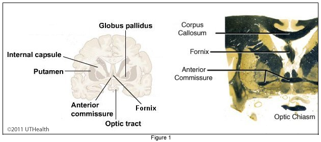
First we’ll consider the fiber tracts: Fornix, Corpus Callosum, Anterior Commissure, and the Optic Chiasm. Next the cell bodies: Amygdala, Uncus and the Parahippocampal Gyrus. Finally, we’ll find the Lateral and Third Ventricles. Recall that fiber tracts appear darker than cell bodies in myelin stains. The following figures are all myelin stains.
The Fornix is a C-shaped structure that is a major input and output pathway of the hippocampus. It begins caudal to the hippocampus and arches superiorly close to the midline under the corpus callosum. It then turns inferiorly and posteriorly toward the hypothalamus.
 The Corpus Callosum, which you saw in the midline view of the wet specimen, joins the two cerebral hemispheres.
At the level of the Internal Capsule Genu we find the Anterior Commissure, a band of fibers that crosses the midline. A large portion of the anterior commissure is responsible for interconnecting the middle and inferior temporal gyri. Notice from the Horizontal section that the anterior commissure has a caudal curve to it. Use this information when you track the anterior commissure in the coronal sections.
Also at the same level, we find the Optic Chiasm. The chiasm is where the fibers of the optic nerve partially decussate to form the optic tracts. Caudal to this level, we see the Optic Tract.
Use the Figure 1 to get a feel for the location of these structures.
Go to the NEXT PAGE