  |
 |
 |
 |
 |
|
|
|
|
|
Lab 01, Page 3 of 38
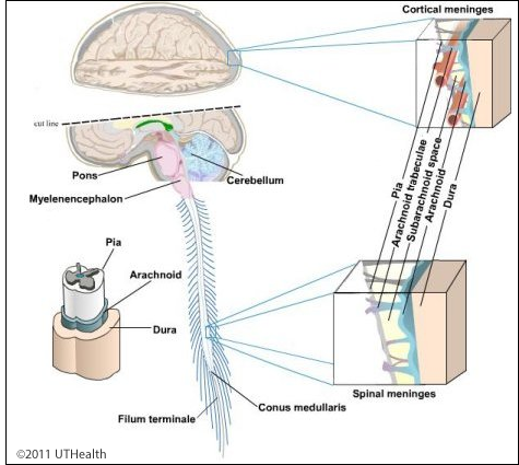 A potential space known as the subdural space separates the dura mater from the delicate arachnoid (spider’s web) below. Like the dura, the arachnoid covers the brain but does not follow the contours of its surface. Contrarily, the innermost pia mater is closely adhered to the surface of the brain following all of its contours. A lattice-work of arachnoid trabeculae extends from the arachnoid to the pia traversing the subarachnoid space, which is normally filled with CSF. In areas where the arachnoid and pia are widely separated, subarachnoid cisterns form. The cerebellomedullary cistern or cisterna magna is located between the base of the cerebellum and the medulla.
If the dura is still present on your specimen, gently raise it from the underlying arachnoid, especially near the longitudinal midline, and note the arachnoid villi (arachnoid granulations) which are extensions of the cerebral arachnoid protruding in the sinuses. In specimens with no dura attached, you may see the arachnoid villi forming small white clusters near the midline ("longitudinal fissure"). The villi, which often become heavily calcified in older adults, serve as one-way valves in passing cerebrospinal fluid into the venous system.
Go to the NEXT PAGE