  |
 |
 |
 |
 |
Lab 9, Page 33 of 42
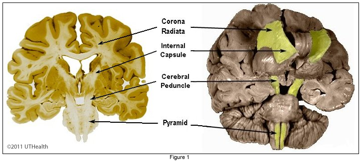
The corticospinal pathway is a one-neuron pathway from the cerebral cortex to the gray of the spinal cord. This pathway consists of all axons that: (1) originate from cells within the cerebral cortex, (2) pass through the pyramids of the medulla, and (3) terminate in the spinal cord. The corticospinal pathway is one of the most prominent descending fiber systems in the human neuraxis. It is concerned with the execution of volitional movements, especially isolated movements of the fingers and hand. Destruction of this pathway results in a deficit in voluntary movement that is most marked in the distal extremities. The proximal joints and grosser movements are less severely and not permanently affected. Lesions involving only the corticospinal path produce deficits, which characteristically are not severe and may, after a time, be difficult to detect.
More content below images.

Most of the cells of origin of this pathway are located in the precentral gyrus (motor cortex), part of the frontal lobe anterior to the precentral gyrus (premotor and supplementary motor area), and parts of the parietal lobe (postcentral gyrus and posterior parietal areas). The axons of these cortical cells converge in the corona radiata, pass downward in the posterior limb of the internal capsule to form the crus cerebri, along with corticobulbar and corticopontine fibers. The corticospinal fibers descend through the pons in compact bundles along with other corticofugal fibers. In the rostral medulla, the corticospinal fibers form a compact bundle, the pyramid, which descends to the caudal medulla where about 90% of the fibers cross in the pyramidal decussation. The crossed fibers form the lateral corticospinal tract of the spinal cord and are located in the posterior portion of the lateral funiculus, medial to the posterior spinocerebellar tract. The uncrossed fibers form the anterior corticospinal tract are located medially in the anterior funiculus along the wall of the anterior median fissure.
The lateral corticospinal tract extends the entire length of the spinal cord and progressively diminishes in size as more and more fibers leave to terminate in the spinal cord gray matter. Below L3, i.e., caudal to where the posterior spinocerebellar tract is found, the lateral corticospinal tract is located along the posterolateral margin of the spinal cord. The fibers of the lateral corticospinal tracts terminate in the ipsilateral cord (i.e., contralateral to the cells of origin).
A smaller portion of the pyramidal fibers descends uncrossed as the anterior or direct corticospinal tract, in the anterior funiculus. Most fibers extend only to the upper thoracic cord. The fibers influence the motor neurons innervating the muscles of the upper extremities and neck. Most of the fibers of the uncrossed anterior corticospinal tract appear to cross in the spinal cord anterior white commissure and to terminate in the contralateral gray. Some remain uncrossed. The contralateral projection (crossed lateral corticospinal tract) influences the neurons innervating distal (chiefly) and proximal limb muscles. The ipsilateral cortical projection (uncrossed anterior corticospinal fibers) influences the motor neurons innervating more proximal limb muscles. About 55% of all corticospinal fibers end in the cervical cord, 20% in the thoracic cord, and 25% in the lumbosacral segments. This suggests that corticospinal control over the upper extremity (hands and fingers) is much greater than over the lower extremity.
Go to the NEXT PAGE