Microscopic Sections of Spinal Cord - Introduction
Lab 4, Page 8 of 32
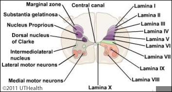
In this part of the lab you will be working with microscopic images of spinal cord sections. Keep the gross spinal cord specimen in mind for comparing external landmarks with internal structures. Work with these images to gain an appreciation of the relative position, shape and size of the nuclear groups and of the different levels of the spinal cord.
More content below images.
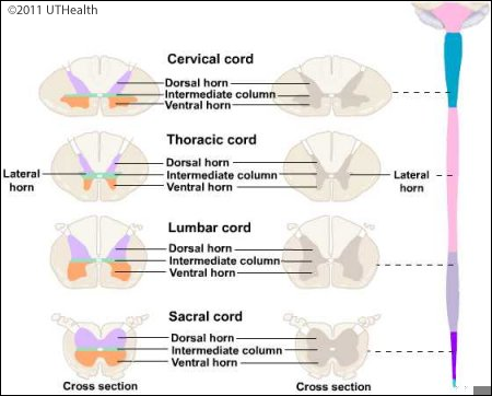
In distinguishing spinal cord levels the following are useful to remember:
- Rostral regions have more white matter because ascending fibers have accumulated from all cord levels and descending fibers have not yet dispersed to their terminations. Conversely, caudal regions have less white matter.
- In regions supplying the limbs and digits (i.e., the cervical & lumbar enlargements), the gray horns are large because they provide a higher density of innervation of skin and muscle. This is because the limbs and digits have increased sensory acuity and discrete motor control.
- The lateral horn is present only at thoracic and lumbar levels.
- The posterior intermediate sulcus subdivides the posterior funiculus at levels above T6.
Go to the NEXT PAGE