  |
 |
 |
 |
 |
|
|
|
|
|
Lab 07, Page 27 of 41
In this section we’ll review the ocular/motor pathways in the pons.
More content below images.
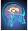
Figure 1, Layer A is approximately at the midpontine level. In the floor of the fourth ventricle, beneath the elevation called the facial colliculus, is the abducens nucleus. Some of its axons, abducens nerve rootlets, can be seen coursing anteriorly where they exit the brain stem at the pons-medullary junction. Recall that the abducens nerve fibers innervate the lateral rectus muscle of the ipsilateral eye. In addition to motor neurons, the abducens nucleus contains abducens interneurons which send ascending axons into the contralateral medial longitudinal fasciculus (MLF) to terminate on oculomotor neurons controlling the medial rectus. At this level, the medial longitudinal fasciculus also contains fibers from the vestibular nuclei that are ascending to the trochlear and oculomotor nuclei.
In the reticular formation near the midline and anterior to the MLF are paramedian pontine reticular formation (PPRF) neurons that constitute the "horizontal gaze center". The "horizontal gaze center" neurons synapse with tectopontine fibers from the superior colliculus and corticofugal fibers from frontal eye field neurons. In turn, these PPRF "horizontal gaze center" neurons send their axons to the ipsilateral abducens nucleus. The axons of abducens nucleus interneurons decussate and ascend in the medial longitudinal fasciculus to the contralateral oculomotor nucleus. This pathway, from the "horizontal gaze center" to the abducens and oculomotor nuclei, is important in the control of saccades (e.g., conjugate horizontal gaze).
Corticofugal fibers terminating in dorsolateral pontine nuclei form part of the circuit controlling smooth pursuit, i.e., eye movements for tracking moving objects. The axons of the pontine nuclei decussate and travel in the middle cerebellar peduncle to the part of the cerebellum associated with the vestibular system. In turn, the cerebellum provides input back to the vestibular nuclei. The vestibular nuclei (superior and medial) are part of the smooth pursuit pathway controlling the movement and trajectory of the eye and send axons via the medial longitudinal fasciculus to the cranial motor nuclei controlling the extraocular muscles.
Figure 1, Layer B: At the level of the caudal pons the abducens nucleus forms a bulge, the facial colliculus, in the floor of the fourth ventricle. The vestibular nuclei occupy the region lateral to the sulcus limitans.
The spinal trigeminal tract and nucleus are located immediately anterior to the vestibular nuclei. They contain the 1° and 2° afferents, respectively, conveying information about somatosensory stimulation of the cornea (as well as other face areas). The facial nucleus is located anteriorly in the pons tegmentum, lateral to the central tegmental tract and posterior to the superior olivary complex. Axons of spinal trigeminal nucleus neurons representing the cornea terminate bilaterally on facial motor neurons innervating the orbicularis oculi of the eyelid. Consequently, somatosensory stimulation of the cornea can elicit a rapid eye blink bilaterally via this eye blink reflex pathway.
Go to the NEXT PAGE