  |
 |
 |
 |
 |
|
|
|
|
|
Lab 5, Page 20 of 26
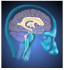
The first set of images are from the brainstem to the diencephalon. Moving to the next section (moving to the right through the images) is moving rostrally. At each level read the corresponding section of text and identify the bold structures.
More content below images.
The posterior/dorsal spinocerebellar tract has been displaced somewhat anteriorly by the spinal trigeminal tract & nucleus and is located posterior to the anterior/ventral spinocerebellar tract. The 1° cuneocerebellar afferents from the upper body are ascending in the cuneate fasciculus to synapse in the lateral cuneate nucleus.
Note that the spinocerebellar tracts have shifted posteriorly and are located along the lateral margin of the posterior medulla. Recall that the posterior/dorsal spinocerebellar tract fibers originate in the dorsal nucleus of Clarke (L3 to C8) and the anterior/ventral spinocerebellar tract fibers originate from the spinal border cells (L5 to T12). Ascending 1° cuneocerebellar afferent fibers carrying information from the upper extremities travel in the cuneate fasciculus, and terminate in the lateral cuneate nucleus. The axons of the lateral cuneate nucleus (2° cuneocerebellar afferents) remain uncrossed (like the axons in the posterior/dorsal spinocerebellar tract) and are collected along the lateral margin of the nucleus in the cuneocerebellar tract.
The posterior/dorsal spinocerebellar tract & the cuneocerebellar tract have entered the inferior cerebellar peduncle and are passing posteriorly to terminate in the cerebellum. The anterior/ventral spinocerebellar tract remains along the lateral margin of the medulla - anterior to the inferior cerebellar peduncle and lateral to the spinal trigeminal nucleus and tract. More rostrally it will join the superior cerebellar peduncle in which it descends to the cerebellum.
The ascending fibers of the anterior/ventral spinocerebellar tract remain along the margin of the medulla lateral to the spinal trigeminal nucleus and tract. Notice the superior cerebellar peduncle in which the descending fibers of the anterior/ventral spinocerebellar tract are traveling to the cerebellum.
The fibers of the anterior/ventral spinocerebellar tract have joined the superior cerebellar peduncle and are beginning their descent to the cerebellum.
Go to the NEXT PAGE